Equine Facilities

  • Digital Radiography
  • Endoscopy
  • Gastroscopy
  • Scintigraphy
  • Surgical Facilities
  • Ultrasonography

Digital Radiography

Radiography (x rays) can be undertaken at our clinic or, using the portable equipment, at home or the livery yard. Generally speaking it is more efficient and cost effective to take x-rays at the clinic, where the images can be developed and viewed immediately. If x-rays are to be taken at home then we need to have access to a reasonably dark covered area, preferably a stable, with a firm level surface and an electricity supply nearby. Generators can be used if absolutely necessary but a mains supply is preferable. The area needs to be quiet and x-raying should normally be arranged for when there are not many people around. The area around the stable will be coned off during the procedure for health and safety reasons and anyone holding the patient must not be pregnant or trying to get pregnant.

Some horses can be x-rayed without the need for any sedation. However, some views require the horse to stand very still or be placed in a slightly uncomfortable position. Also, we are sometimes working in a confined area. For these reasons your vet may decide to give a light sedative which will make the whole process smoother and safer for everyone including the patient.

If the x-rays are taken at the clinic you will normally be shown the images as soon as they have been developed. If they are taken at home then your vet would normally call you later in the day or the following day to discuss the findings. Although the hard copies remain the property of the practice, you are always welcome to view the images or to have them forwarded to another practice if you leave the area.

In the near future we plan to invest further in radiographic services by purchasing state of the art digital radiography. This technology enables us to obtain exceptional quality images allowing the earlier detection of abnormalities and injuries. Appropriate interventions and treatments can then be started at an earlier stage in the disease process, hopefully improving the response to treatment, resulting in a better long term prognosis in many cases.

Endoscopy

Endoscopy is primarily used to examine the respiratory tract from the nostrils to the lungs including the sinuses, pharynx (back of throat), guttural pouches, larynx, trachea and lungs. It can also be used to examine the back of the mouth, the oesophagus (food pipe) and stomach, along with the reproductive tract, bladder and rectum.

We have recently purchased a new endoscope with video and still image recording capabilities, allowing us to record examinations and view the procedure on a monitor rather than having to peer down the end of the scope. The extended length of our new scope allows complete examination of the stomach to identify any evidence of gastric ulceration.

The endoscopy equipment includes a 2.7m long cable connected to a camera, which is in turn connected to a laptop computer where the image is viewed. Still photos and video footage are stored on the computer for future reference. Endoscopy can be performed either at the clinic or at home. It requires a dark stable with a power supply nearby. Depending on the area being examined your vet may need to sedate the patient lightly as some horses/ponies will not tolerate the procedure.

Gastroscopy

Gastroscopy is a procedure that we can readily perform at our clinic to examine a horse's stomach. It involves passing a fibre optic camera up a horse's nose, down the oesophagus and then into the stomach. To perform this the horse must have been starved for at least 10 hours, and not been allowed to drink for the hour prior to the procedure, commonly they are sedated for this procedure.

During this procedure we can examine the oesophagus, the lining of the stomach and where the small intestine leaves the stomach, including where bile is ejected to mix with the food.

We may recommend this procedure if we suspect the horse has gastric ulcers, is displaying signs of recurrent colic or weight loss, or has had an episode of choke and we need to check the oesophagus has not become damaged. We can also perform biopsies of the start of the small intestine this way, a part called the duodenum.

Scintigraphy

Nuclear Scintigraphy (Bone Scanning)

We designed a purpose built scintigraphy room and installed a gamma camera as part of our facilities at the Brooke Equine Clinic. This is housed alongside two stables specifically as an isolation unit. 

Since its installation scintigraphy has proved very useful, in particular:

  • In horses or ponies with multilimb lameness for which a work up would prove extremely lengthy and difficult
  • Suspected fracture cases as a bone scan is extremely sensitive for these and can pick up hairline fractures where an X-ray may not
  • Horses with suspected dental problems. Bone scan will show sites that are currently actively inflamed
  • To image parts of the body that are difficult to investigate and X-ray. For example the pelvis and hips
  • Animals not suited to using needles in their legs!

A labeled radioactive isotope – Techenetium99 is injected into the horse. This attaches to sites where bone is remodelling; where bone metabolism is increased there will be an increased amount of isotope attached. The camera picks up these areas of increased bone turnover as increased radioactive uptake or ‘hot spots’. 

If your horse is having a Bone Scan: 

We like to have the horse in the day before to allow it to settle in and so that we can start early the following morning. Please bring a few thick rugs and 4 stable bandages and gamgee as we rug and stable bandage the horse overnight to ensure good circulation. 

On the day of the bone scan horses are lunged in our arena until warm, fully rugged and bandaged, and then have an intravenous catheter inserted in to the jugular vein. 

In cases where lunging is not possible they stand under some heat lamps until warm, this ensures good circulation to all parts of the body, enabling the best possible images. 

Once injected with the isotope, the horse remains in the stable for 2 hours prior to starting scanning. 

Although a safe procedure, to comply with radiation regulations horses are kept in an isolation stable and allowed to go home approximately 60 hours after injection, or when they are sufficiently un-radioactive.

Surgical Facilities

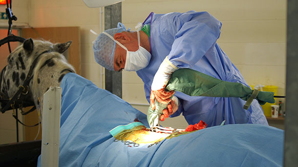
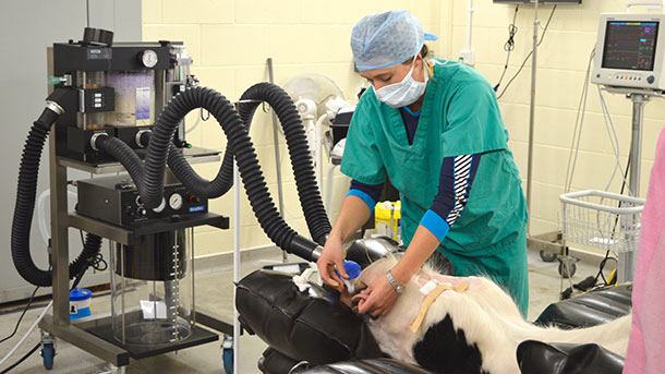
We are very fortunate to have purpose built surgical facilities where we can undertake general anaesthetics on equine patients ranging in size from the day old foal to fully grown heavy horses. The electric hoist system and operating table allow the safe positioning of patients and the specially padded recovery room provides an area for horses to recover during the dangerous recovery period with minimum risk of injury.

All operations involve at least one equine surgeon, one equine anaesthetist, and one or more equine nurses. All our equine surgical staff have many years of combined experience, undertaking the delicate task of anaesthetizing equine patients as safe as possible, using modern equipment, drugs and surgical procedures to minimize the risk of complications.

Elective Surgery

The majority of elective (non-life threatening) procedures undertaken involve the treatment of orthopaedic injuries. We also undertake a range of other procedures involving the head/neck such a dental/sinus surgery and involving the abdominal tissues such as hernia repairs.

If your horse requires an elective surgical procedure this will either be undertaken using standing sedation and local anaesthesia, or it will be necessary to give the patient a full general anaesthetic. In either case you will be asked to bring the patient to the clinic either first thing in the morning on the day of the procedure or in the afternoon the day before. If they are coming in on the morning of the procedure, usually between 8-9am, you will need to remove all access to hard feed and hay/grass about 12 hours before hand. Water can be provided until you load the patient to bring them to the clinic. If they come in the day before then we will starve them overnight. You do not need to bring any food with you unless they have any specific requirement or supplements. Otherwise, you will simply bring a head collar, lead rope and any rugs that they would normally wear, including a sweat rug for use immediately after surgery when the patients are frequently quite sweaty.

A nurse will ask you to read and sign a consent form on admission and you will be contacted by either the nurse or the surgeon once the patient has stood up after a general anaesthetic or after a standing procedure is complete. The period of stay after surgery depends on the procedure being undertaken, but your vet should be able to tell you approximately how long this will be once the procedure is completed.

Arthroscopy (Keyhole Surgery)

We frequently use keyhole surgery to treat a range of injuries and conditions of equine joints and tendon sheaths, both as elective and emergency procedures. Arthroscopic surgery allows minimally invasive examination and treatment of conditions such as OCD, chip fractures and infected joints/tendon sheaths, allowing accurate assessment and treatment of injured tissues, with small wounds and resultant shortened recovery times.

Emergency Surgery

We also have the facility to undertake emergency surgery, normally including emergency colic surgery and treatment of wounds such as joint penetrations, fractures or tendon lacerations. If we are not able to undertake the procedure at our clinic we will arrange for immediate referral to a surgical centre, usually Rossdales Equine Hospital in Newmarket.

Whether the patient is undergoing elective or emergency surgery, it is important that the issue of finances is discussed with the owner, if possible before the procedure is undertaken. Please see the section on insurance claims for further details on how to initiate a claim for the cost of veterinary fees.

The vast majority of owners are aware of the high cost of surgical procedures on equine patients. If the patient is insured for the cost of veterinary fees then the decision of whether or not to operate is normally easier to make, although it is very important to check the limits and restrictions of any policy at the earliest opportunity, clarifying the level of cover with the provider if necessary. It is our experience that clients much prefer to know the sorts of costs likely to be incurred beforehand. Whilst it is often difficult to provide an accurate cost estimate due to the unpredictable factors inherent in equine surgery, we are happy to discuss costs and, if requested, provide a price range estimate. We also feel that, while discussing finances when faced with the need for emergency surgery, agreeing terms of payment at any early stage in the treatment causes considerably less distress for everyone further down the line. Please feel free to discuss this unavoidably sensitive area at any time with your veterinary surgeon or one of our administrative staff.

Ultrasonography

Ultrasonography is a non-painful and non-invasive means of assessing external and internal soft tissues and the surfaces of bone/joints.

We currently have three ultrasound machines. Two are portable and are primarily used for fertility work during the breeding season. With these machines we can monitor changes in the reproductive tract when mares are cycling to help with natural breeding. We also frequently scan mares before, during and after undertaking artificial insemination (see AI section for more information). We also use these machines to investigate reproductive and urinary tract problems. To carry out an ultrasound exam at home we need a stable or reasonably dark covered area with a power supply nearby. One of our machines also works off a portable battery if no power supply is available, although we would need to know this in advance so that the vet brings the correct machine. Leg scans can also be carried out using one of the portable machines, however the image quality is inferior to the clinic based scanner.

Our clinic based digital ultrasound machine has greatly enhanced our ability to investigate leg swellings/injuries, abdominal problems, heart and eye abnormalities, creating exceptional quality images using extremely sensitive ultrasound probes. The main benefit of this machine is that it allows us to identify much milder injuries or damage to structures at an earlier stage than would be possible using the portable equipment, thereby preventing more serious damage to tissues like tendons and ligaments. The result is that treatment can be started sooner and the prognosis will be significantly better than had the injury been detected at a later stage. Although the use of this machine requires a trip to the clinic, our clients almost always feel the trip was worthwhile.

Return to Equine